
Ethos Therapy System & Ion Robotic-Assisted Bronchoscope
All across Chester County Hospital, there are examples of how state-of-the-art technology is redefining medicine. We're highlighting two potentially groundbreaking innovations in this issue. The artificial intelligence-driven Ethos therapy system is enabling our radiation oncologists to personalize treatment plans to an unprecedented degree, in real time. Meanwhile, our pulmonologists are biopsying nodules in parts of the lungs that were previously largely unreachable for such a procedure.
This is made possible by the Ion robotic-assisted bronchoscope, a breakthrough that could change the way the deadliest of all cancers is detected. With these additions comes new hope: for more treatment options, for improved quality of life, and for life after disease.
State-Of-The-Art Technology in Radiation Oncology
>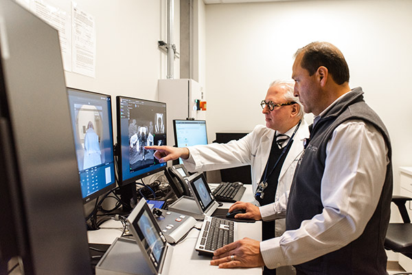
Radiation Oncologist Andre A. Konski, MD utilizing The Ethos Therapy System's artificial intelligence technology.
State-of-the-art technology is enabling Chester County Hospital's radiation oncologists to modify treatment plans on a day-to-day basis and target tumors more precisely than ever.
State-of-the-art, artificial intelligence-driven technology is helping radiation oncologists at Chester County Hospital personalize cancer care to an unprecedented degree. And it appears that’s just scratching the surface of how AI could revolutionize healthcare in the near future.
ChatGPT captured the world's imagination over the last year. But the use of chatbots, specifically, and AI, in general, has already become fairly commonplace in medical settings across the United States. It's being used to transcribe medical documents, improve physician-patient communication, and, increasingly, to help diagnose patients and assist doctors in identifying treatment protocols, clinical tools, and appropriate drugs more efficiently.
The Ethos Therapy System, introduced last spring in the Penn Medicine Radiation Oncology Program at Chester County Hospital, falls under the latter categorization. It’s considered an adaptive therapy, which means that, unlike traditional methods, Ethos provides the ability to modify a treatment plan based on changes to the tumor and the patient’s anatomy. By enabling doctors to better visualize these often-subtle differences, the system allows radiation oncologists to be more accurate with each dose they deliver and more precise with their delivery.
The ability to alter and adapt radiotherapy treatments has long been the aspiration of radiation oncologists. That’s because the human body is not a static entity, particularly during cancer treatment, says Andre A. Konski, MD, MBA, MA, FACR, FASTRO, Medical Director, Radiation Oncology, Chester County Hospital.
Currently, Ethos is being used at the hospital to treat patients with pelvic malignancies.
"When we do our treatment planning, we assume the bladder and rectum are going to be in the same position every day, and they’re just not," Dr. Konski says. "There are going to be differences in how full the bladder and bowel are from day to day. And those changes can have a big impact on the treatment."
Neighboring organs and healthy tissue can also shift. And, ideally, the tumor will shrink over the course of treatment. Yet conventional treatment plans were largely unable to account for this, at least not on a daily basis. But Ethos therapy easily integrates various high-quality diagnostic images – MRI, PET, CT – as well as daily iterative cone beam computed tomography images at the console within a matter of minutes. With a detailed, real-time view of the patient’s anatomy enabled by Ethos, the radiation oncologist can adapt the prescribed therapy accordingly. This quickly produces several customized plans, showing a variety of possible radiation dose distributions.
"We can see what’s changed with their anatomy and then make slight adjustments to the original treatment plan," Dr. Konski says. "Each day, then, we’re reassessing the appropriate dose of radiation and how to deliver it most effectively."
As cancer treatments have become more innovative, they've also become more potent and complex. Radiotherapy patients are now frequently treated with higher daily doses. And cancer patients routinely receive other cancer therapy simultaneously, potentially exposing them to higher risks for treatment-related side effects. Adjusting the dosage and how it's delivered at each treatment could help reduce patients' side effects, which, for the types of cancer being treated with Ethos, could include skin irritation, fatigue, nausea, sexual dysfunction, and bowel and urinary problems.
Dr. Konski says it's too early to predict how Ethos will impact overall outcomes. To advance adoption of the system, Varian, the company that developed it, has formed a consortium and is encouraging radiation oncologists around the world to collaborate and share their data. The consortium is expected to lead clinical trials with the aim of developing evidence-based clinical protocols for Ethos. At Chester County Hospital, Dr. Konski says the system may be used to treat a broader selection of cancer patients in the coming months if it continues to display advantages over traditional practices. But he can't yet say how pronounced those advantages are.
This much he can say with certainty: AI is here to stay in healthcare.
Beyond the limited adoption of Ethos, AI is also being used in the Radiation Oncology program to help generate the contours of healthy tissue, essentially providing doctors with a clearer view of the tumor.
"Almost every patient who has curative cancer will have treatment that allows us to use AI to help generate the normal tissues," Dr. Konski says. "As AI learns more, it's going to be used in more and more applications. From what we've seen so far, it’s promising, but it still needs to be vetted and studied over the long term."
Innovation Opens Access To The Lungs (and Not a Moment Too Soon)
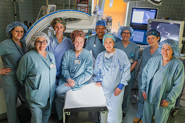
Pulmonologist Rajesh J. Patel, MD and Team
A new, minimally invasive bronchoscopy is changing the way lung cancer is detected. Such a breakthrough has been long awaited, with lung cancer often not diagnosed until the later, deadlier stages of the disease.
A groundbreaking, robotic-assisted bronchoscope that was introduced at Chester County Hospital last June appears to have the potential to significantly impact the leading cause of cancer deaths in both women and men in the United States.
Lung cancer is more deadly than breast cancer, prostate cancer, and colon cancer combined, according to Michael R. Costello, MD, Medical Director of the Abramson Cancer Center.
"The reason is that 75 percent of lung cancer cases are diagnosed at an advanced stage," Dr. Costello says. "While there are very good treatments now available, lung cancer in the later stages, unfortunately, is still not curable."
Those considered at high risk of developing lung cancer are not being screened at anywhere near the rate of those who are at risk of developing breast cancer, which is a missed opportunity for early detection, says Rajesh J. Patel, MD, FCCP, a pulmonologist at Chester County Hospital.
The difference is reflected in the five-year survival rates. For men with prostate cancer that hasn't spread, it's 100 percent. For women with non-metastatic invasive breast cancer, it's 91 percent. For all types of lung cancer, it's 23 percent. But if lung cancer is caught before it spreads, the five-year survival rate improves to 63 percent, according to the American Lung Association.
The earlier the diagnosis, the more treatment options there are available, Dr. Patel says. But the issue is more nuanced than a lack of screening.
"A lot of lung cancers begin as these small, peripheral nodules that traditional bronchoscopies aren't able to reach," Dr. Costello says.
Dr. Patel estimates that 70 percent of lung cancer nodules are located in the outer third of the lung, a "hard-to-reach area full of tight spaces and narrow airways. "In other words, even if imaging done during a screening was to detect a suspicious shadow, biopsying that nodule has posed such a serious challenge, historically, that the only practical option in many instances was to wait and see if the growth became larger.
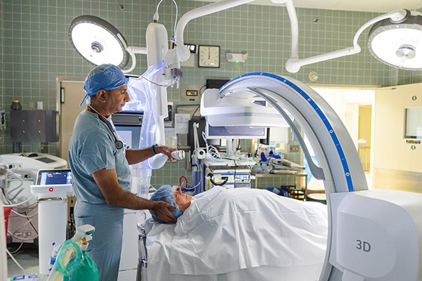
But that is changing with the Intuitive Ion robotic-assisted bronchoscope. Now, a pulmonologist can extract a tissue sample as small as one centimeter from parts of the lung not accessible to standard lung biopsy methods.
Using a controller, the pulmonologist guides an ultra-thin robotic catheter along a preplanned route to the nodule. CT scan data is used to generate a three-dimensional image of the patient’s lungs down to the branches and nodules. The highly maneuverable catheter is able to navigate tight turns. Once it reaches the destination, the catheter locks onto the nodule and a needle obtains the tissue sample.
"With Ion, we're able to perform biopsies on peripheral lung lesions earlier than we previously could," Dr. Patel says. This, combined with a higher rate of lung cancer screenings, "has the potential to be transformative," Dr. Costello says.
"One of The More Impressive Advances I've Seen"
Prior to the arrival of Ion, which was also implemented last year at the Abramson Cancer Center at Pennsylvania Hospital, another option for biopsying peripheral lung nodules was a procedure called a transthoracic needle biopsy. Here, an interventional radiologist inserts a needle between the patient's ribs and into the lung using ultrasound or CT scans, which guides them to the nodule.
The procedure is not without risk. The needle makes a small hole in the lung, which will usually seal itself. If it doesn't, air can leak out into the space around the lung causing the lung to collapse. This happens in about one in four people, according to the American Thoracic Society.
Reaching the nodule and collecting enough tissue for a biopsy are also not givens. The smaller the nodule, the more challenging its location is to reach, and the greater the degree of difficulty.
While transthoracic needle biopsies are performed at Chester County Hospital, they do not have "the same level of success with securing a diagnosis" as the Ion endoscope, according to Erin Kini, Director of Operations for Procedural Services at the hospital.
In planning to replace the hospital's existing endoscope, she says she originally budgeted for one that was half the cost of Ion, but there were concerns from the hospital's radiologists that its technology was becoming outdated. It also was incompatible with their newer, state-of-the-art imaging equipment.
Kini says what ultimately compelled her to commit to the higher cost was understanding "that we could expand our offerings to people who would otherwise only be monitored. With this technology, we'd be able to go after their cancer sooner. At that point, investing in Ion became imperative."
The early results create a striking picture. Of those referred to Chester County Hospital between last June and December because of a suspicious finding on a CT scan of their lungs, 90 percent received a definitive diagnosis. (During this period, 27 people underwent an Ion bronchoscopy at the hospital.) Before the Ion bronchoscope was introduced, 50 percent of referrals received a definitive diagnosis.
"When you talk about increasing a diagnostic yield from 50 to 90 percent, that's a massive improvement," Kini says. "We're bringing new patients into the fold. Previously, the standard was let's wait and watch because the nodule's too small, and we know we're not going to have success biopsying it. That we're now giving people an option they didn't have before makes Ion one of the more impressive advances I've seen recently."
What To Expect and Why It's Empowering
Pulmonologist Gaurav J. Patel, MD
Most lung nodules are benign. The only way to know that for certain is through a biopsy, which entails collecting a tissue sample from the suspicious area and examining the extracted cells under a microscope.
The type of biopsy procedure your doctor may recommend will depend on the size of the nodule and its location within the lung. If they suggest a bronchoscopy with Ion, they'll refer you to one of Chester County Hospital's two pulmonologists, Rajesh J. Patel, MD, FCCP, and Gaurav J. Patel, MD.
The outpatient procedure is performed in an operating room. Dr. Rajesh Patel says that multiple tissue samples are collected from the nodule, as well as the nearby glands. One or two slides are sent immediately to a pathologist, who will then provide a diagnosis within 10 minutes. After the procedure, he'll send additional slides to the pathologist. This time, they'll do a more involved type of analysis that could take up to five days before they confirm the diagnosis.
If lung cancer is detected, a staging workup is the next step, because pinpointing a timeline for the disease is critical for determining the treatment options. This includes an MRI (magnetic resonance imaging) of the brain and a PET (positron emission tomography) scan.
There are two types of lung cancer. Small-cell carcinoma typically spreads more quickly and is generally more aggressive than non-small-cell carcinoma, so they have different stages.
The prospect of either can be concerning. But beyond the initial shock, there's empowerment.
"The sooner you know there's a problem," says Kini, a former operating room nurse, "the higher the chance it can be addressed with less aggressive means and the greater the likelihood you'll have a good outcome."
At the Abramson Cancer Center's three convenient locations in West Chester*, Kennett Square*, and Exton*, PA, patients have access to cancer infusion services in a healing environment and a team of hematology/oncology providers who specialize in the prevention, early detection, diagnosis, and treatment of cancer.
Together, our world-class, team of oncologists, specialists and researchers work to create the world's most advanced cancer therapies.
*A Facility of the Hospital of the University of Pennsylvania