You’re watching your favorite hospital show on TV. The patient has a mysterious illness. As the doctors try to figure out the diagnosis, they tack images of the inside of the patient’s body to a backlit wall and pace back and forth.
This type of scene isn’t very accurate, especially since most images are viewed on a computer screen, rather than on a wall.
What Are Imaging Tests?
Imaging tests allow providers to see inside your body and how it functions. Sometimes these tests are used during a procedure, but when they are used to diagnose or monitor a condition, it’s called diagnostic radiology.
For some tests, your provider may give you a contrast solution — a temporary substance that makes it easier to see certain body parts. This can be given by mouth or through an IV.

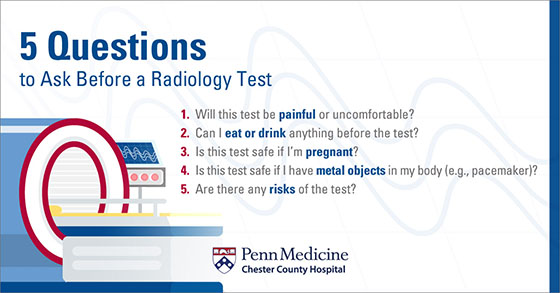
While many radiology tests are safe for pregnant women, there are some additional risks. Your physician may recommend certain accommodations or waiting until you’ve given birth to get the test.
Here are 4 of the most common diagnostic radiology tests.
1. X-rays
X-rays are the oldest, most commonly used form of medical imaging.
You will receive a very small dose of electromagnetic radiation, which travels through your body to produce black and white pictures.
Why You Might Need an X-ray
X-rays produce some of the clearest and most detailed views of bones, so they’re frequently used to diagnose fractures and spine or joint injuries. They can also be used to:
- Look for arthritis or growths on your bones
- Locate foreign objects in your body that you may have accidentally swallowed
- Assist in detecting and diagnosing bone cancer
X-rays in Mammograms
Mammograms — tests to screen for signs of breast cancer — also use X-rays. Many mammograms are now 3D mammograms. They take multiple X-rays and put them together to make a 3D image of the breast. These are more detailed than regular mammograms, and also give a better image of dense breast tissue.
3D mammograms have improved cancer detection rates by 41% and have reduced the number of times a woman has to come back for additional imaging by 15%.
Getting an X-ray
Getting an X-ray is quick and painless. It usually takes less than 15 minutes. Even though X-rays use radiation, they are very safe, especially if you wear protective gear.
2. Computed Tomography (CT)
Computed tomography (CT), or CAT scans, are like advanced X-rays. A CT machine takes X-rays from multiple angles. The images — called slices — are assembled to create detailed 3D pictures.
CT scans also use radiation, but in controlled amounts.
Why You Might Need a CT Scan
X-ray images show bones very clearly, but not muscles, tendons, or joints. CT scans, on the other hand, show breaks, injuries, and tears to your bones, muscles, tendons, and joints.
Common uses of CT scans include:
- Spotting tumors, including cancerous tumors in the abdomen, pelvis, chest, lung, liver, kidneys, pancreas, or ovaries
- Detecting blood clots, particularly ones that could cause a stroke
- Finding excess fluid in the lungs
- Diagnosing breathing conditions, such as pneumonia or emphysema
- Examining injuries
Getting a CT Scan
During the scan, you will lie down on a table that moves through a structure shaped like a donut, called a gantry. As the table moves, X-ray beams rotate around you to take pictures. The machine can look a little foreboding, but don’t worry — like X-rays, CT scans are totally painless.
Results are usually available within 2 to 3 days, but they’re often available sooner.
+ A PET Scan
Positron emission tomography (PET) scans are often taken right alongside CT scans. They measure body functions, such as bloodflow, oxygen use, or sugar use, to show how well your organs and tissues are working.
Together, CT and PET scans provide an even more specific picture of what's happening inside your body.
3. Magnetic Resonance Imaging (MRI)
An MRI uses a powerful magnetic field, radio waves, and computer software to produce highly detailed images of the organs and structures in your body. They do not use any radiation.
Why You Might Need an MRI
MRIs provide more detail about your body than X-rays and CT scans. They’re particularly useful for finding problems in the brain, spinal cord, and nerves — such as epilepsy or multiple sclerosis. MRIs are also good at detecting signs of cancer.
Additionally, MRIs can be used to:
- Identify problems with reproductive organs, such as the uterus or prostate
- Evaluate joint problems, such as tissue tears and sprains
- Diagnose or monitor conditions like congenital heart disease or inflammatory bowel disease
- Provide images of how blood flows through your arteries and veins (called magnetic resonance angiography, or MRA)
Getting an MRI
You will lie down on a table that slides into a tube in the MRI scanner, and the technologist will take a series of pictures. It’s important to lie completely still during this so they can get the images.
MRIs take a little longer than some of the other imaging tests — anywhere from 15 minutes to an hour.
Since scanners tend to make loud banging noises, you will be given headphones or earplugs. You may be able to listen to music to pass the time.
Results can take a couple of days, but you may be able to get them sooner.
But … Small Spaces
If you’re claustrophobic (scared of enclosed spaces), you might be worried about a test where you’re enclosed in a tube.
However, there are a few reasons why you don’t need to worry:
- You will be given a button — usually in the form of a squeeze ball — to use if you feel panicked or like you need to get out. You can press it at any time.
- The technologist will communicate with you throughout the test. Between each series of pictures, they will make sure you’re doing okay and let you know how long the next series of pictures will take.
- In some cases, the technologist may be able to ask your provider to prescribe a sedative to help you stay calm
**At Chester County Hospital, we offer a “Try it On For Size” program where you can visit the MRI units and get any of your questions answered on the spot. Call 610-431-5131 for more information or to schedule a tour.**
4. Ultrasounds
Ultrasounds use sound waves to see internal organs and blood flow. Ultrasounds don’t use radiation.
Why You Might Need an Ultrasound
Pregnant women receive ultrasounds to confirm a pregnancy and monitor the baby’s growth and development, including any signs of birth defects or complications.
An ultrasound can also examine organs throughout your entire body, including your heart, liver, kidneys, bladder, gallbladder, and reproductive organs. It’s a great test to help your provider determine what’s causing pain, swelling, or infection.
Getting an Ultrasound
The provider will move a device called a transducer around the area of your body being studied. The transducer captures the sound waves and sends them to a machine that creates the images.
Ultrasounds generally don’t hurt. If you are receiving an ultrasound because you’re pregnant, the transducer may be inserted directly into your vagina, which can be a bit uncomfortable.
It’s normal to be a little nervous about any test. But remember that just because your provider orders a test, it doesn’t mean that something is necessarily wrong. And if a test does show a problem, that’s the first step toward getting the treatment you need.
Additional Radiology Information from Chester County Hospital:
There are many other types of imaging tests. Read more about these and other tests available at Chester County Hospital.
Also Read:
Scheduling a Radiology Test at Chester County Hospital
You will need a referral for most radiology tests. For more information about imaging tests, call 610-431-5130. To schedule an appointment for any test (except PET scans), call 610-431-5131. To schedule a PET scan appointment, call 610-495-0060.